Look Inside Your Body (Look Inside Board Books): 1
FREE Shipping
Look Inside Your Body (Look Inside Board Books): 1
- Brand: Unbranded

Description
In 1973, American chemist Paul Lauterbur (1929–2007) showed that NMR could produce images. British scientist Peter Mansfield (1933–2017) developed the mathematical processes that turned MRI into a useful rapid imaging technique. Lauterbur and Mansfield were awarded the Nobel Prize in Medicine in 2003. Dr Singh says: "So many of us tend to put off seeing our doctors, or simply deny that there’s anything wrong. Confronting our conditions, face-to-face with these incredible 3D images, means there’s nowhere to hide. And that can prompt patients to ask really difficult questions. Armed with more knowledge about their own bodies, I saw patients ask not just about their treatment and recovery, but also about life death and everything in between. The power of this technology is that it allows doctors and patients to talk about the stuff that really matters." Peek under all the flaps in these colorful and engaging books–perfect for little fingers and curious minds.”– Usborne Look Inside Your Body Inside Look Inside Your Body R Bud and D J Warner (eds), Instruments of Science, An Historical Encyclopaedia (London: Science Museum, 1998) J Bronzino, V Smith and M Wade (eds), Medical technology and society: an interdisciplinary perspective (Massachusetts: MIT press 1990)
Look Inside Your Body PaperPie. Look Inside Your Body
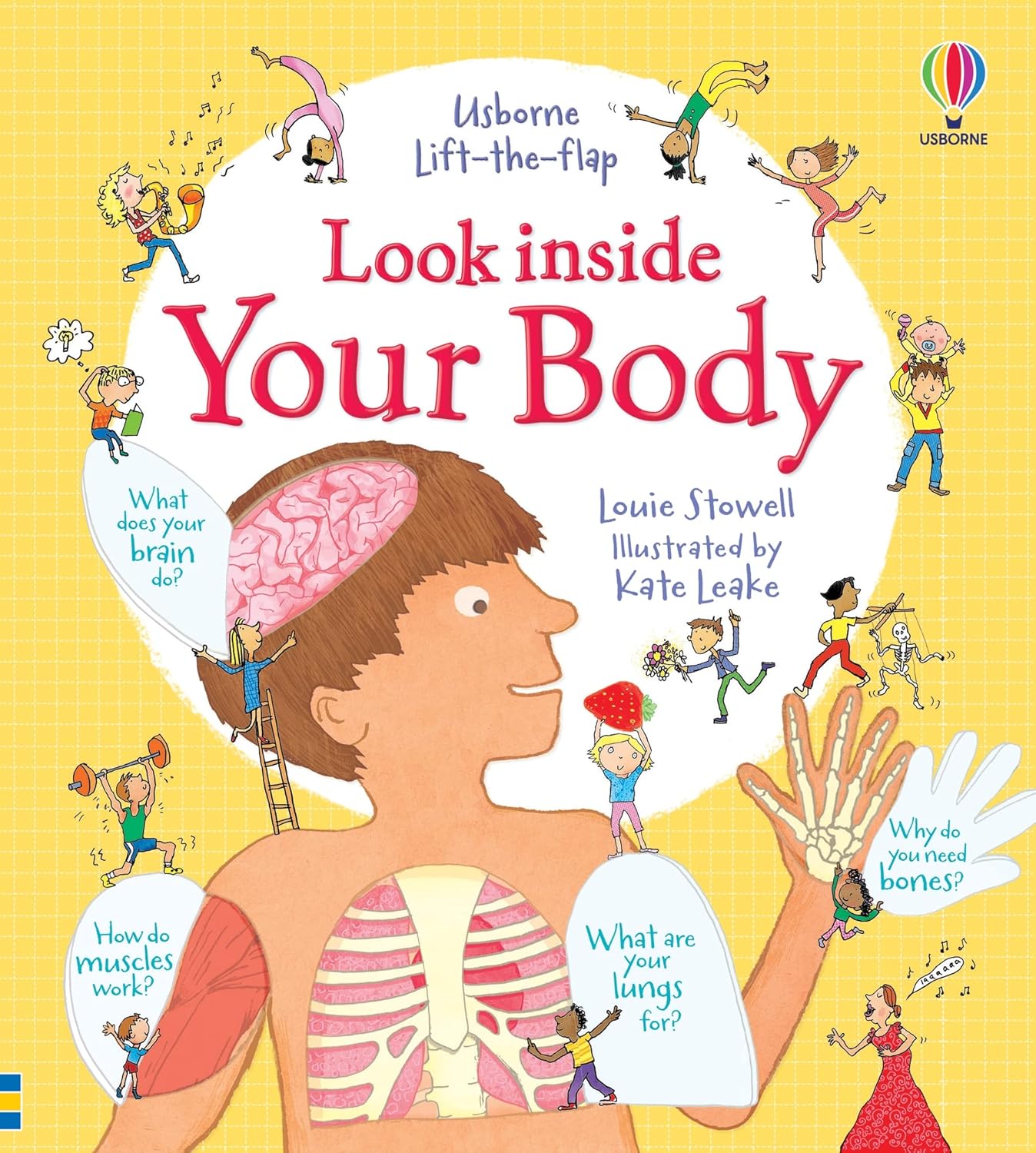
It is written in an informative, factual but informal way which is beneficial because it is adding to the children’s vocabulary as well as understanding things that are going on with their own bodies in a fun way. W F Bynum and R Porter (eds), Companion Encyclopedia of the History of Medicine (London: Routledge, 1993) E Koch, ‘In the image of science?: Negotiating the development of diagnostic ultrasound in the cultures of surgery and radiology’, Technology and culture, 34 (1993), pp 858-893 The book is all about the human body from the digestive system to the skeleton and everything in between. There is a lot of detail but not too much that makes it difficult for the children to read. The technology was impressive, particularly when they compared the size of Hilda’s uterus to a normal one. Gynaecologist Mr Stephen Quinn told her it would be wise to operate, though warned that if there was too much blood loss, he might have to remove her uterus entirely. Despite wanting to have children, Hilda recognised the importance of having the operation and Mr Quinn ended up removing an incredible 100 fibroids. Dr Dimitri Amiras, Trudi and Kate Garraway looking at a GFX representation of Trudi’s frozen shoulder (Photo: BBC/Remarkable TV)
However, PET is not used in medicine as often as other scanning techniques. PET techniques are complex and expensive, partly because they require enormous machines called cyclotrons to produce the radioactive tracers.
Visualising the body | Science Museum
Dr Singh says: "The 3D images of the patients’ bodies are incredible. As a doctor, it sometimes feels that language isn’t up to the job of truly explaining what is happening inside the gloriously complex human body. How amazing, then, to have a tool that allows realistic, accurate and personal images to do some of the job for you! When you’re ill, the first step towards recovery is coming to terms with your diagnosis, and I think being able to see, literally, what’s going on inside your own body gives patients the understanding to really do that. But this isn’t just for patients – the truth is, despite all my years of medical training, I’d never seen the human body quite like I did on this show. Being on this show has been a real first for me. I’ve never experienced health in this way, and it’s been an eye-opener." R B Gunderman, X-ray Vision: The Evolution of Medical Imaging and its Human Significance (Oxford: Oxford University Press, 2013) Because MRI can construct images of soft tissue, it's especially useful for diagnosing joint abnormalities, diseases of the liver and abdominal organs, and identifying tumours and uterine conditions such as fibroids.
Dr Singh says: "There have been so many, not least the look on all the patients’ faces when they first came face-to-face with their own bodies in larger-than-life technicolor. But in the first episode we meet Hilda, who had one of the worst cases of fibroids her consultant had ever seen. My mouth literally dropped when we were treated to augmented reality image of what it would look like to have all her 90-plus fibroids lined up in a row. But even more astounding was just how different Hilda seemed when I met her after her operation to remove them. The physical transformation alone was mind-boggling, but what brought tears to my eyes was just how much more alive and whole she seemed, too. She was a different woman!" In 1971, American scientist Raymond Damadian (1936–) discovered that MRI could be used for medical diagnosis. The radio signals emitted by cancer cells in a tumour were different from those in healthy cells and could be isolated by the MRI scanner. Damadian built the first whole-body MRI scanner in 1977, which he called the 'Indomitable'. Ultrasound is a diagnostic imaging technology that uses high-frequency sound waves—well beyond the range of human hearing—to produce pictures of the inside of the body. In MRI, the patient is placed in a powerful magnetic field, which influences the hydrogen atoms in the body. Short bursts of radio waves are then used to alter the atomic alignment created by the magnetic field. When the radio waves are turned off, the atoms return to their alignment and in so doing emit a weak radio signal of their own. CT scans provide more detailed images than X-ray machines. They can be used to detect bone and joint damage, including complex bone fractures. They can also reveal the precise location, size and shape of unusual occurrences such as tumours and blood clots, as well as internal injuries such as bleeding.
Look Inside Your Body - Louie Stowell | PDF - Scribd Look Inside Your Body - Louie Stowell | PDF - Scribd
B Holtzmann-Kevles, Naked to the Bone: Medical Imaging in the Twentieth Century (New Jersey: Rutgers University Press, 1997) An MRI scanner uses magnetic fields and radio waves to generate images of the inside of the body. Unlike X-rays, an MRI scan can visualise soft tissue such as the organs and blood vessels. It is a safe and painless procedure, leaving no lasting effect on the patient. The writing is split up into small bubbles of writing and the children are able to work their way round the book in a creative way. Having the flaps in the book also add that element of excitement making it a fun learning tool. K A Joyce, Magnetic Appeal: MRI and the Myth of Transparency (Ithaca, N.Y.: Cornell University Press, 2008) It would be useful throughout KS1-2. Even if the younger children are not fully aware of all the terms they are still able to use their fine motor skills when using the flaps etc.Learn more about your body in this lift-the-flap Look Inside Your Body Usborne book. Flaps are layered under flaps to dive deeper into the body layers. Ultrasound scanners were not commonly used in hospitals until the 1970s. By the 1980s the technology had advanced enough to produce moving images in shades of grey, followed by 3D imaging not long after. Today ultrasound is widely used in surgical procedures and the field of gynaecology. Yet I was left with more questions than answers. Are these methods used in real-life diagnosis? If not, why not? Why had these women been allowed to live in such pain for so long? More interrogation into the whys and wherefores would have been appreciated. An MRI scanner detects these weak signals. Because each of the body’s tissue types emits a different frequency of radio waves, the MRI scanner can distinguish between them and build an image based on the data it receives.
- Fruugo ID: 258392218-563234582
- EAN: 764486781913
-
Sold by: Fruugo